
Intranodal palisaded myofibroblastoma: Difference between revisions
SimLibrarian (talk | contribs) m fix missing space |
m Disambiguating links to Spindle cell (disambiguation) (link changed to Spindle neuron) using DisamAssist. Tag: Reverted |
||
| Line 33: | Line 33: | ||
==Diagnosis== |
==Diagnosis== |
||
IPMs are diagnosed by examination of the [[Tissue (biology)|tissue]] by a [[pathologist]].{{cn|date=February 2021}} |
IPMs are diagnosed by examination of the [[Tissue (biology)|tissue]] by a [[pathologist]].{{cn|date=February 2021}} |
||
They have a rim of peripheral [[lymphoid tissue]] (remnant of a [[lymph node]]) and consist of [[spindle cell]]s with [[cell nucleus|nuclear]] [[palisade|palisading]]. [[Red blood cell]] [[extravasation]] is common and [[blood vessel]]s surrounded by [[collagen]] with (fine) peripheral [[spoke]]s (amianthoid fibers) are usually seen.<ref name=pmid1918406>{{Cite journal | last1 = Bigotti | first1 = G. | last2 = Coli | first2 = A. | last3 = Mottolese | first3 = M. | last4 = Di Filippo | first4 = F. | title = Selective location of palisaded myofibroblastoma with amianthoid fibres. | journal = J Clin Pathol | volume = 44 | issue = 9 | pages = 761–4 |date=Sep 1991 | doi = 10.1136/jcp.44.9.761| pmid = 1918406 | pmc=496726}}</ref> |
They have a rim of peripheral [[lymphoid tissue]] (remnant of a [[lymph node]]) and consist of [[Spindle neuron|spindle cell]]s with [[cell nucleus|nuclear]] [[palisade|palisading]]. [[Red blood cell]] [[extravasation]] is common and [[blood vessel]]s surrounded by [[collagen]] with (fine) peripheral [[spoke]]s (amianthoid fibers) are usually seen.<ref name=pmid1918406>{{Cite journal | last1 = Bigotti | first1 = G. | last2 = Coli | first2 = A. | last3 = Mottolese | first3 = M. | last4 = Di Filippo | first4 = F. | title = Selective location of palisaded myofibroblastoma with amianthoid fibres. | journal = J Clin Pathol | volume = 44 | issue = 9 | pages = 761–4 |date=Sep 1991 | doi = 10.1136/jcp.44.9.761| pmid = 1918406 | pmc=496726}}</ref> |
||
[[Immunostain]]s for [[smooth muscle]] [[actin]] and [[cyclin D1]] are characteristically positive. The main [[histologic]] [[differential diagnosis]] is [[schwannoma]].{{cn|date=February 2021}} |
[[Immunostain]]s for [[smooth muscle]] [[actin]] and [[cyclin D1]] are characteristically positive. The main [[histologic]] [[differential diagnosis]] is [[schwannoma]].{{cn|date=February 2021}} |
||
Revision as of 18:22, 29 April 2024
| Intranodal palisaded myofibroblastoma | |
|---|---|
 | |
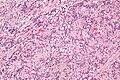
| Micrograph of an intranodal palisaded myofibroblastoma. H&E stain. | |
| Specialty | Oncology |
Intranodal palisaded myofibroblastoma (IPM) is a rare primary tumour of lymph nodes, that classically presents as an inguinal mass.[1]
It afflicts predominantly males of middle age.
Signs and symptoms
IPMs present as painless lymphadenopathy.[1] They usually are found in the inguinal region and grow slowly. The signs and symptoms are non-specific, i.e. it is not possible to diagnose an IPM from the symptoms and manner in which they present.[citation needed] The main (clinical) differential diagnosis of IPM is metastatic cancer, e.g. squamous cell carcinoma, malignant melanoma, adenocarcinoma.[citation needed]
Diagnosis
IPMs are diagnosed by examination of the tissue by a pathologist.[citation needed] They have a rim of peripheral lymphoid tissue (remnant of a lymph node) and consist of spindle cells with nuclear palisading. Red blood cell extravasation is common and blood vessels surrounded by collagen with (fine) peripheral spokes (amianthoid fibers) are usually seen.[2]
Immunostains for smooth muscle actin and cyclin D1 are characteristically positive. The main histologic differential diagnosis is schwannoma.[citation needed]
-
 Low mag.
 -
 High mag.


Treatment
Simple surgical excision is considered curative. Rare recurrences have been reported.[3]
See also
References
- ^ a b Nguyen, T.; Eltorky, MA. (Feb 2007). "Intranodal palisaded myofibroblastoma". Arch Pathol Lab Med. 131 (2): 306–10. doi:10.5858/2007-131-306-IPM. PMID 17284119.
- ^ Bigotti, G.; Coli, A.; Mottolese, M.; Di Filippo, F. (Sep 1991). "Selective location of palisaded myofibroblastoma with amianthoid fibres". J Clin Pathol. 44 (9): 761–4. doi:10.1136/jcp.44.9.761. PMC 496726. PMID 1918406.
- ^ Creager, AJ.; Garwacki, CP. (May 1999). "Recurrent intranodal palisaded myofibroblastoma with metaplastic bone formation". Arch Pathol Lab Med. 123 (5): 433–6. doi:10.5858/1999-123-0433-RIPMWM. PMID 10235504.